Research


Computer-aided detection of early Barrett's cancer in Endoscopic Video
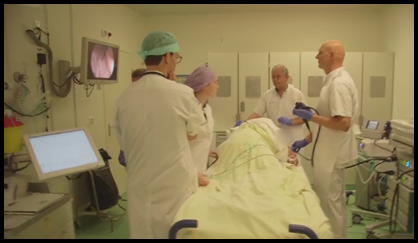
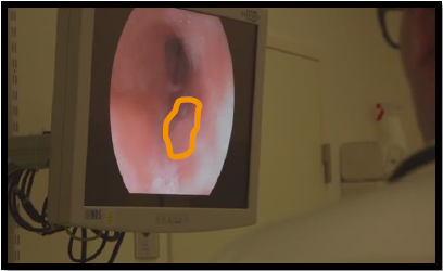
Early cancerous lesions in Barrett's Esophagus (BE) are very hard to detect and are often overlooked during endoscopic surveillance of the esophagus. When the disease is detected early, the cancerous tissue can be surgically removed by endoscopic mucosal resection. This treatment knows very high curation rates, showing 93% of the patients in complete remission after 10 years. However, when a lesion is missed, and the disease is detected in a late stage, the prognosis is grave with a five-year survival rate of approximately 15%, hence it is crucial that these cancerous lesions are identified at an early stage. Therefore, we aim at developing Computer-Aided-Detection algorithms, that support the gastroenterologist in detecting these early lesions during endoscopic surveillance.
Additional material:
 Set of slides with the basic concepts (dutch).
- More comprehensive overview of the study (as of december 2015).
Consortium:

Automated analysis of Volumetric Laser Endomicroscopy (VLE) scans
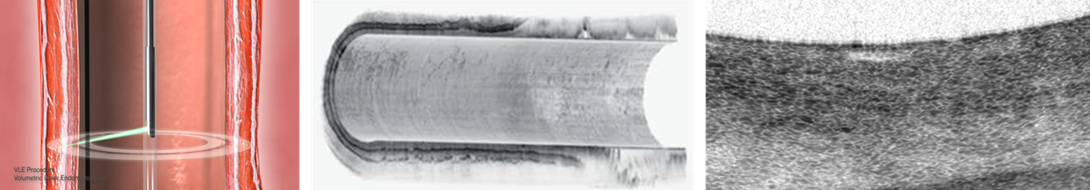
Volumetric Laser Endomicroscopy (VLE) is a recent endoscopic imaging technique that allows gastroenterologists to inspect the submucosal tissue layers of the esophageal wall. This is a very useful imaging property for the detection of early esophageal cancer, which typically develops in these subsurface layers that cannot be imaged by traditional endoscopic imaging techniques. During a VLE image capturing procedure, a balloon is endoscopically inserted in the esophagus. Next, the balloon is inflated to stretch the tissue into a fixed position after which a laser scans a cylindrical segment of up to 6 cm in 90 seconds. Althoug VLE is fast, efficient and scans an entire segment of the esophagus up to a depth of 3 mm, there are two major difficulties that this novel technique faces: (1) physicians struggle to interpret the complex VLE signal and (2) carefully inspecting 1200 high-resolution VLE slices is very laborious and time consuming. Therefore, we aim to develop a Computer-Aided Detection (CADe) algorithm that automatically inspects a full VLE volume for signs of early cancer.
Consortium:

Depth estimation of surgical instruments for Image Guided Interventions
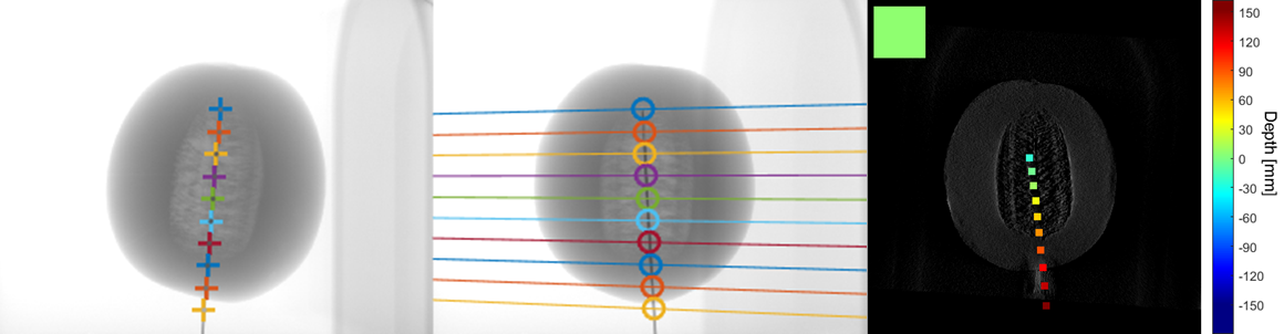
For many image-guided biopsy procedures, a needle is employed for extracting a tissue sample. Since the physician cannot see the needle directly, X-ray imaging can be used to visualize the position of the needle. Typically, the C-arm is placed in a fixed position for this procedure, yielding a two-dimensional projection of the volume. This makes real-time 3D alignment of the biopsy needle and a target nodule impossible. An alternative approach is to record a full scan when the needle is close and employ tomosynthesis to reconstruct the volume. However, this process is quite cumbersome, since medical staff has to move away from the rotating C-arm and the position information of the needle loses its accuracy as soon as the physicians continue the biopsy procedure. Methods for obtaining depth information of a surgical needle without interrupting the intervention are highly attractive. During the PANORAMA project we have investigated a novel approach for depth estimation of biopsy needles, in which we split the problem in two parts -- needle depth estimation and volume reconstruction -- which enables solving them independently.
Additional material:
- Set of slides with the basic concepts.
Consortium: